
Bioimaging
Nuclear microscopy is the use of nuclear microprobes for the purposes of imaging. [1] This includes techniques like scanning transmission imaging microscopy (STIM), ion induced fluorescence (IF) [2, 3], proton induced X-ray emission (PIXE) and Rutherford backscattering (RBS). [4]
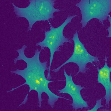
STIM image of HeLa cells
Here we show an example of scanning transmission imaging microscopy (STIM) images obtained with the CIBA single cell imaging facility [5] where we are routinely capable of obtaining beam spot sizes < 50 nm for imaging, greatly surpassing the diffraction limit of conventional optical techniques.
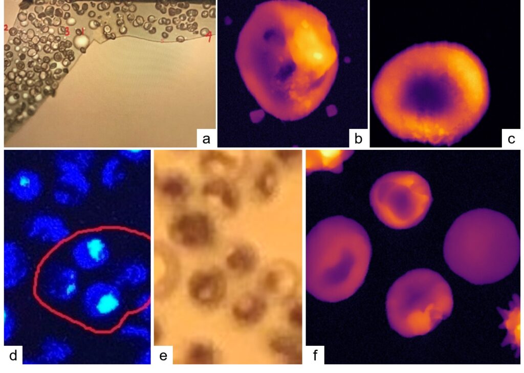
STIM images of red blood cells fixed on 100 nm thick silicon nitride window. The brighter regions in a STIM image correspond to regions of higher areal density. The top row (a,b,c) are optical and STIM images, the cells in (b) and (c) are labelled in the optical image (a) as 3 and 4 respectively. The STIM image sizes are 7.4 by 7.4 µm. The second row (d,e,f) are fluorescence, optical and STIM images respectively.
The region in the STIM image is circled in red on the fluorescence image. The STIM image is 15 by 15 µm. With STIM we can obtain high resolution information of the cell interior as the ions are able to pass through the whole cell without much deviation, unlike electrons used in TEM or SEM.
References
| [1] | M.Q. Ren, X Ji, S.K. Vajandar, Z.H. Mi, A. Hoi, T Walczyk, J.A. van Kan, A.A. Bettiol, F. Watt, T. Osipowicz, Analytical possibilities of highly focused ion beams in biomedical field, Nuclear Instruments and Methods in Physics Research Section B: Beam Interactions with Materials and Atoms, Volume 406, Part A, 2017, Pages 15-24. |
| [2] | Andrew A. Bettiol et al, Ion beam induced fluorescence imaging in biological systems, Nuclear Instruments and Methods in Physics Research Section B: Beam Interactions with Materials and Atoms, Volume 348, 2015, Pages 131-136. |
| [3] | Mi, Z., Chen, CB., Tan, H.Q. et al. Quantifying nanodiamonds biodistribution in whole cells with correlative iono-nanoscopy. Nat Commun 12, 4657 (2021). |
| [4] | M. Ren, Reshmi Rajendran, Mary Ng, Chammika Udalagama, Anna E. Rodrigues, Frank Watt, Andrew Michael Jenner, Nuclear microscopy of rat colon epithelial cells, Nuclear Instruments and Methods in Physics Research Section B: Beam Interactions with Materials and Atoms, Volume 269, Issue 20, 2011, Pages 2264-2268. |
| [5] | F. Watt et al, The Singapore high resolution single cell imaging facility, Nuclear Instruments and Methods in Physics Research Section B: Beam Interactions with Materials and Atoms, Volume 269, Issue 20, 2011, Pages 2168-2174. |
Content by Frederick Cheong, 2023